INTRODUCTION
As of January 1, 2026, the Council on Accreditation of Nurse Anesthesia Educational Programs (COA) requires that all nurse anesthesia residents receive formal instruction in point-of-care ultrasound (POCUS) as part of their training.1 The COA defines POCUS as “the use of portable ultrasonography at a patient’s bedside for diagnostic purposes,” and specifies that the use of ultrasound solely for image-guided procedures (for example, vascular access or regional anesthesia) does not satisfy this diagnostic POCUS requirement.1
This mandate has created an urgent curricular challenge for many nurse anesthesia programs, which must quickly integrate diagnostic ultrasound scanning into existing training. Emerging data highlight the scope of the challenge: a recent survey of nurse anesthesia faculty found that 10% of respondents reported POCUS was not part of their formal curriculum, and only 5–19% of faculty self-identified as competent in gastric scanning or transthoracic echocardiography (TTE).2 A principal barrier is the limited number of faculty who are trained and confident in performing and teaching diagnostic POCUS.2
Given these constraints, programs should adopt evidence-based educational strategies to develop POCUS competency efficiently and reliably. The literature demonstrates that POCUS skills are teachable across learner groups, including nurse anesthesia residents and medical students, and that multimodal instructional approaches produce the best outcomes.3–5 Effective POCUS training typically combines didactic instruction, supervised live scanning, and simulation.6–8
POCUS competency is not purely knowledge-based; current definitions in the literature encompass a range of cognitive and technical skills. Competent performance includes9,10:
-
Recognizing the clinical indications for the examination
-
Applied knowledge of ultrasound equipment
-
Image acquisition and optimization
-
Conducting a focused examination using a systematic approach
-
Accurate interpretation of findings
-
Appropriate documentation of the examination
-
Clinical decision-making informed by exam findings
Because many of these competencies require hands-on practice, particularly image acquisition and focused exam performance, didactic lectures alone are insufficient.7,8 Supervised live scanning with models or peers is essential for teaching probe handling, image orientation, and recognition of normal anatomy, and it closely approximates scanning a human subject.7 However, live scanning has inherent limitations: volunteer or standardized patients are typically healthy and rarely present the critical pathologies nurse anesthetists must recognize, including severe aortic stenosis, cardiogenic shock, or tamponade. In addition, standardized patients incur associated costs, incidental abnormal findings may lead to unnecessary follow-up, and learners may experience increased anxiety when practicing on live models.11
Low-fidelity static phantom manikins address some practical constraints but are of limited educational value because they typically represent normal anatomy and offer little realistic pathology.9 High-fidelity, dynamic simulation can reproduce clinically significant, time-sensitive pathophysiology and provide repeated, stress-free hands-on practice.9 Such simulation fills an important gap: while didactic teaching conveys the necessary knowledge and live scanning teaches normal examinations, few programs offer opportunities for learners to perform focused, hands-on scanning of patients with acute, complex physiologic derangements in a controlled learning environment. Addressing this gap is essential to meeting the COA POCUS content requirement and ensuring nurse anesthesia graduates are prepared to apply diagnostic POCUS in perioperative and critical care settings.
METHODS
A POCUS simulator is a training device that recreates ultrasound scanning and interpretation within a controlled environment.7,9 POCUS simulators can be delivered in multiple different formats including virtual, and computer-based platforms that use a plug-in probe device to interact with on-screen ultrasound images (for example, SonoSim).9 Other simulators are manikin-based, where learners scan a physical manikin to generate ultrasound images that correspond to probe position and orientation (for example, BodyWorks Eve from Surgical Science).9 Both formats aim to provide realistic, repeatable practice opportunities, though manikin systems may offer a greater sense of spatial realism.9
The manikin-based POCUS simulator pairs a realistic, ergonomically designed probe and transducer interface with software-driven, high-fidelity ultrasound images and physiologic models so learners can practice image acquisition, optimization, focused exam protocols, and diagnostic interpretation without the need for a live patient.9 These systems allow hand–eye coordination training by linking probe position and orientation to real-time image generation on an anatomical manikin or virtual patient. These devices also often include haptic feedback to approximate the feel of an actual transducer.9
Typical POCUS manikin-based simulators provide a library of programmed patient cases that span normal anatomy and a range of pathologies across cardiac, pulmonary, abdominal, and vascular domains, with adjustable physiologic parameters to simulate dynamic clinical changes. They offer guided learning modes, such as stepwise tutorials, checklists, and competency assessments, alongside free-scan practice for skill consolidation, and record performance metrics (image quality, correct views obtained, timing) to support objective assessment and remediation. Because they enable repeatable, low-risk practice and exposure to high-acuity, time-sensitive pathologies that are uncommon in live models, POCUS simulators serve as valuable adjuncts for developing both the technical and interpretive skills required for clinical competence.7,9
Educational Frameworks
The I-AIM Framework
The I-AIM framework is a structured, stepwise approach to performing and teaching point-of-care ultrasound examinations. Originally described by Bahner and colleagues12 and subsequently applied across multiple POCUS applications,13 I-AIM provides a procedure-specific, standardized method that improves consistency, supports rapid and accurate image acquisition, and links sonographic findings directly to clinical action.12,13 The acronym represents 4 sequential steps12,13:
-
Indication- The clinician identifies the specific clinical question or reason for performing the ultrasound examination. In perioperative and critical care settings, common indications include hemodynamic instability, respiratory distress, or assessment of volume status.
-
Acquisition-The clinician obtains the ultrasound images using proper patient positioning, probe selection (e.g., phased-array for cardiac imaging), scanning technique, and optimization of ultrasound settings (depth, gain, focus). This step emphasizes systematic, protocol-driven scanning to ensure examination consistency and image quality.
-
Interpretation- The clinician analyzes the acquired images using pattern recognition to identify normal anatomy versus pathologic findings. Interpretation requires integration of sonographic appearance with clinical context to generate a differential diagnosis.
-
Medical decision-making (Management)- Based on the interpreted findings, the clinician determines the appropriate clinical intervention. This may include changes to anesthetic technique, timing of surgery, hemodynamic management, or escalation of care.
By organizing POCUS instruction around the I-AIM framework, learners develop a reproducible cognitive approach that links the technical act of scanning to clinically meaningful decisions.12,13 This structure is well-suited to simulation-based practice where scenarios can be designed to require explicit progression through each step.
The FATE Examination
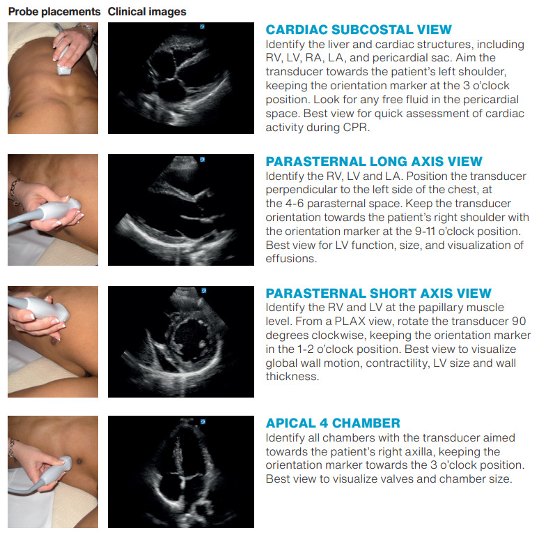
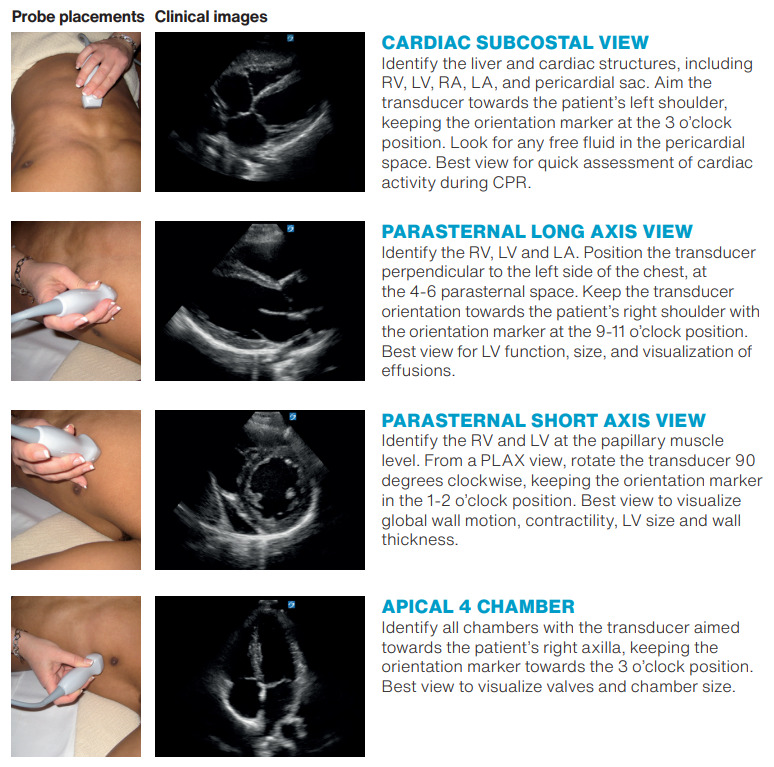
The Focus Assessed Transthoracic Echocardiography (FATE) examination is a validated, goal-directed cardiac ultrasound protocol developed for perioperative and critical care settings.14,15 FATE is designed to answer focused clinical questions rapidly at the bedside rather than provide a comprehensive echocardiographic study. The examination comprises 4 standard transthoracic cardiac views plus assessment of bilateral pleural spaces15:
-
Subcostal 4-chamber view- Allows visualization of all 4 cardiac chambers, pericardial space, and assessment of global cardiac function; often the easiest view to obtain in supine, mechanically ventilated patients.
-
Apical 4-chamber view- Provides comparison of right and left ventricular size and function, assessment of valvular motion, and evaluation for pericardial effusion.
-
Parasternal long-axis view- Enables evaluation of left ventricular size and contractility, aortic and mitral valve structure, left atrial size, and pericardial space.
-
Parasternal short-axis view- Allows assessment of left ventricular wall motion by coronary artery territory, ventricular septal position (indicating right ventricular pressure/volume overload), and global contractility.
-
Bilateral pleural assessment- Identifies pleural effusions, which may contribute to respiratory compromise or suggest underlying pathology.
In the perioperative setting, this focused scanning protocol is high-yield because it provides timely, actionable information relevant to a wide range of pathophysiologic states and anesthesia emergencies, including hypovolemia, left ventricular failure, right ventricular strain, cardiac tamponade, and severe valvular disease.14 The FATE examination can typically be completed in minutes and does not require advanced echocardiographic training, making it well-suited as a foundational competency for nurse anesthesia residents.
Application to Simulation-Based Education
Organizing simulator sessions around the I-AIM framework and the FATE examination protocol allows faculty to structure learning objectives explicitly. During each simulated scenario, students are expected to: (1) articulate the indication for imaging based on the clinical stem provided; (2) acquire the standard FATE views systematically while optimizing image quality; (3) interpret findings by comparing acquired images to mental models of normal and pathologic anatomy; and (4) state how identified abnormalities would inform immediate anesthetic or resuscitative management. Faculty-led debriefing reinforces this stepwise reasoning and provides targeted feedback on technical acquisition, interpretive accuracy, and clinical decision-making.
Application of the Intervention
At a university-based school of nursing in the Mid-Atlantic United States, nurse anesthesia residents receive POCUS instruction via didactic lectures, live scanning labs with standardized patients, and hands-on time with a POCUS manikin-based simulator. For a simulator, this program specifically uses the CAE Vimedix (CAE, Montreal, Quebec, Canada). Teaching POCUS is organized around the I-AIM framework,12,13 and curricular emphasis is placed on achieving competency in the FATE examination (Figure 1).15 Simulation sessions are conducted in small groups of 4-5 learners with 1 faculty instructor, permitting focused supervision and individualized feedback.
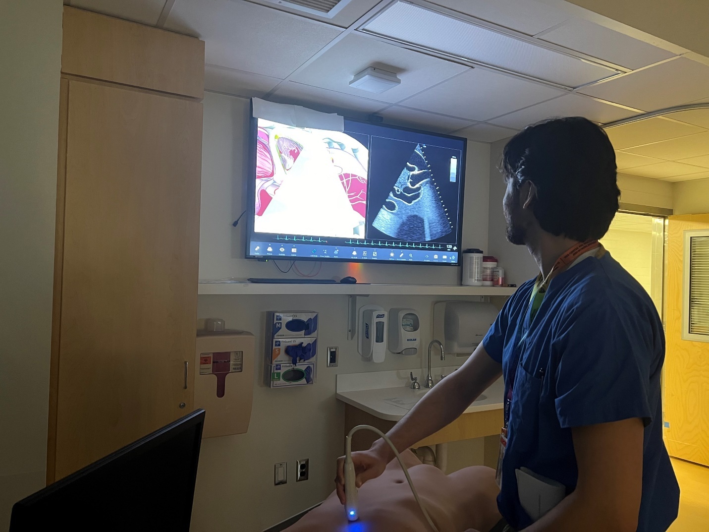
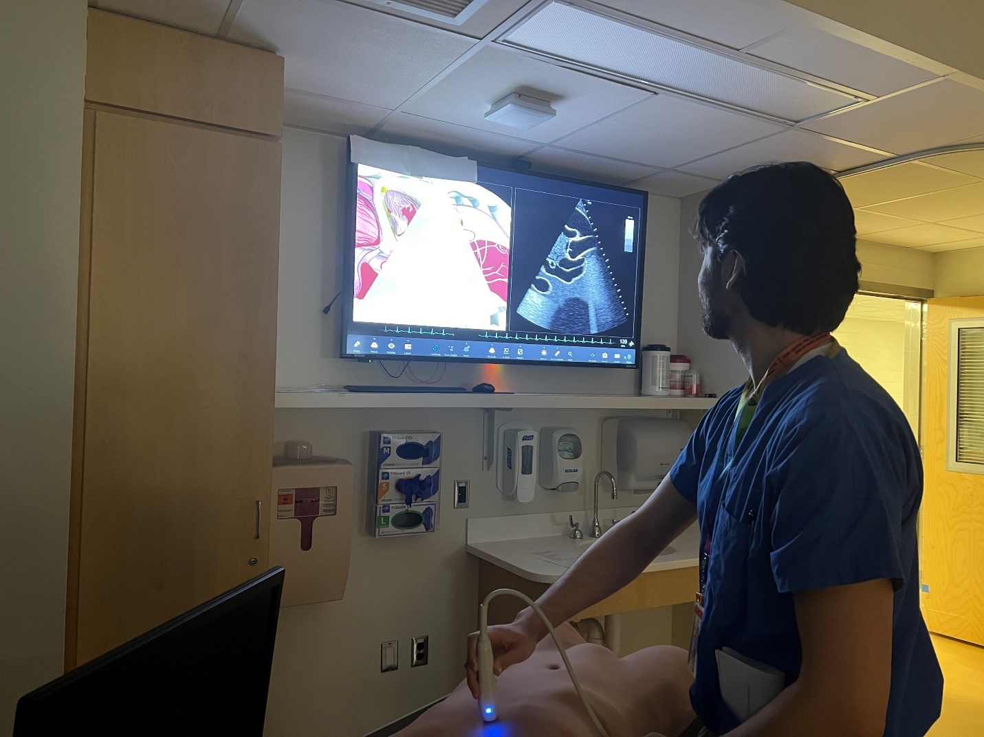
The CAE Vimedix POCUS simulator integrates a physical transducer interface, a lifelike anatomical manikin or scan pad, and advanced software that generates high-fidelity, real-time ultrasound images linked to probe position and orientation; the system plugs into a desktop or laptop computer on which learners view both the ultrasound and corresponding anatomical images. Spatial-tracking technology detects probe movement and translates it into corresponding cross-sectional images drawn from a library of patient cases and physiologic models, permitting learners to acquire and optimize views as they would on a real patient.9 The manikin accepts multiple probes, typically linear, curvilinear, and phased-array transducers. The manikin also typically includes a transesophageal echocardiography probe, enabling practice across a broad range of examinations and applications. Image rendering supports adjustable ultrasound controls (depth, gain, focus, and Doppler functions) and dynamic physiologic parameters (heart rate, rhythm, chamber size, valve function, and hemodynamics) so pathologic states appear and evolve realistically. The platform integrates directly with Vimedix software, which allows instructors to script or change clinical scenarios, conceal or reveal specific pathologies, and modify ultrasound settings in real time; built-in tutorials and objective performance metrics found on some simulator models support deliberate practice, assessment, and curricular integration.9
Each simulation session lasts approximately 90 minutes and is structured to allow every student repeated hands-on practice. Learners alternate between scanning normal anatomy and encountering programmed pathophysiologic conditions, thereby developing both routine image-acquisition skills and the ability to recognize abnormal findings. The simulator’s case library and instructor-controlled scenarios permit reproducible exposure to a range of cardiac, pulmonary, and hemodynamic abnormalities that are infrequently observed in live models.
We use the following pre-programmed pathophysiologic cardiac conditions using the CAE Vimedix POCUS Simulator:
-
Severe left ventricular systolic dysfunction (cardiogenic shock pattern)- reduced global contractility, dilated left ventricle, low ejection fraction.
-
Right ventricular dilation and strain (acute right ventricular failure / massive pulmonary embolism pattern)- right ventricular enlargement, McConnell’s sign, septal flattening.
-
Cardiac tamponade- pericardial effusion with diastolic right atrial/ventricular collapse and respiratory variation in inflow.
-
Severe aortic stenosis- concentric left ventricle hypertrophy with low stroke volume and characteristic hemodynamic consequences; Doppler evaluation if available.
-
Hypertrophic obstructive cardiomyopathy (dynamic left ventricular outflow tract obstruction)- asymmetric septal hypertrophy, systolic anterior motion of the mitral valve, and dynamic gradients.
-
Regional wall motion abnormality (acute ischemia/infarction)- focal hypokinesis or akinesis in coronary distribution.
-
Severe hypovolemia (low preload)- small, collapsible left ventricle and hyperdynamic heart with reduced cavity size; inferior vena cava small and highly collapsible if included.
Clinical realism is introduced through case-based pre-briefing and concealed instructor programming of the manikin’s physiologic state. For example, a learner may be presented with a brief clinical stem, such as “a 62-year-old patient with an intraoperative blood pressure drop to 64/33 mmHg,” and asked to describe anticipated assessment steps and anesthesia interventions before scanning. The student then performs a FATE examination on the manikin while blinded to the specific programmed pathology, simulating the diagnostic uncertainty present in real clinical encounters.
During scanning, students are encouraged to pose focused clinical questions that guide their POCUS assessment and narrow the differential diagnosis. They are taught to confirm any abnormal finding in more than 1 ultrasound view before using it to guide clinical management.Top of FormBottom of Form The clinical questions include:
-
Is the cardiac image normal or abnormal?
-
Is there a pericardial effusion? (would be seen as an anechoic strip around the heart)16
-
Are the ventricles squeezing well? (indicates contraction / ejection fraction)16
-
Are the valves opening and closing? (indicates clinically significant valvular stenosis / regurgitation)16
-
Are the atria and ventricles relatively normal in size? (indicates ventricular or atrial dilation)16
-
Is the myocardium thicker or thinner than normal? (indicates hypertrophy / dilated cardiomyopathy)16
-
Is the right ventricle bigger than the left ventricle? (possibly indicates pulmonary embolus / pulmonary hypertension)16
-
Is the whole left ventricle squeezing? (indicates regional wall motion abnormalities which could be significant for an acute myocardial infarction)16
Throughout the encounter, the I-AIM framework structures learner behavior: students must articulate the indication for imaging, detail the planned acquisition strategy (views, probe position, and ultrasound settings), interpret images in real time including a differential diagnosis, and state how findings would inform immediate medical decision-making.12,13 Faculty-led debriefing follows each scenario, focusing on technical image acquisition, interpretive accuracy, and clinical management decisions, with targeted feedback to guide subsequent practice and consolidate competency.
Evaluation of Learner Perceptions
The Simulation Effectiveness Tool–Modified (SET-M)
After integrating the POCUS simulator into the competency-based POCUS curriculum in 2023, the investigator assessed learner perceptions using an adapted version of the Simulation Effectiveness Tool–Modified (SET-M).17 The SET-M is a validated revision of the original Simulation Effectiveness Tool (SET), which was developed for the Program for Nursing Curriculum Integration. Leighton et al17 subsequently updated the instrument to reflect current simulation standards and established the psychometric validity and reliability of the revised SET-M. The standard SET-M contains 19 items organized into 3 subscales that evaluate distinct phases of the simulation experience: 1.) Prebriefing (2 items assessing whether prebriefing increased confidence and was beneficial to learning), 2.) Scenario (13 items addressing preparation for patient changes, understanding of pathophysiology, confidence in assessment skills, empowerment in clinical decision-making, understanding of medications, opportunity to practice clinical decision-making, confidence in prioritizing care, confidence in patient communication, confidence in patient teaching, confidence in reporting to the healthcare team, confidence in patient safety interventions, and confidence in evidence-based practice), and 3.) Debriefing (5 items evaluating contribution to learning, opportunity to communicate feelings, value in improving clinical judgment, opportunities for self-reflection, and constructive evaluation of the simulation). Each item is rated on a Likert-type scale, with higher scores indicating greater perceived effectiveness.17
Modifications for This Activity
For this study, the survey instrument was modified in 2 ways to align with the specific educational context. First, 3 custom items were developed to evaluate the POCUS simulator station specifically, using the same response format as the SET-M17:
-
“This station contributed to my learning.”
-
“This station will help me in clinical practice.”
-
“The feedback from this station was valuable.”
These items were accompanied by an open-ended prompt: “Please provide any additional feedback regarding the POCUS practice session (on the POCUS simulator).”
Second, the standard SET-M scenario items were adapted by adding the stem “After the POCUS practice session (on the POCUS simulator)…” to each item (e.g., " After the POCUS practice session (on the POCUS simulator), I am better prepared to respond to changes in my patient’s condition") to contextualize responses. The complete survey also included parallel sets of items evaluating the POCUS lecture and live scanning workshop components of the curriculum, as well as a global item assessing whether respondents would recommend the POCUS module. Although the survey evaluated multiple components of the curriculum, the analyses presented in this manuscript are limited to the 3 quantitative items and qualitative responses related to the POCUS simulator station.
RESULTS
All students who participated in the simulator-based POCUS education were invited to complete an anonymous electronic survey administered via Qualtrics (Qualtrics, Provo, UT). Survey invitations were distributed following completion of the POCUS module, and participation was voluntary. Responses were collected over 2 consecutive student cohorts across 2 years, for a total of 48 students who received the survey. Twenty-five students completed the survey, yielding an overall response rate of 52% (25/48). This study was reviewed by the Johns Hopkins University School of Medicine Institutional Review Board and determined to be exempt from full review, as it involved analysis of de-identified student data. For analysis, descriptive statistics were performed on each survey item, and qualitative responses were reviewed for recurring themes.
Quantitative Results
Table 1 presents the complete distribution of responses to the 3 simulator-specific items. Overall, learner perceptions were highly positive. For the item “This station contributed to my learning,” 23 respondents (92%) selected “Strongly agree” or “Agree,” with 1 respondent (4%) selecting “Disagree” and 1 respondent (4%) not providing a response. For “This station will help me in clinical practice” and “The feedback from this station was valuable,” 24 respondents (96%) selected “Strongly agree” or “Agree,” with 1 respondent (4%) not providing a response for each item.
Qualitative Feedback
Open-ended comments specifically addressing the POCUS simulator were uniformly positive (Table 2). Representative comments included “The simulator was very helpful” and “Seeing different pathology was helpful.” No negative qualitative comments were received regarding the simulator station itself.
DISCUSSION
The findings of this study demonstrate strong learner acceptance and perceived effectiveness of a simulation-based POCUS curriculum utilizing the CAE Vimedix simulator, the I-AIM framework, and the FATE examination protocol for nurse anesthesia residents. With 92-96% of respondents indicating strong agreement or agreement regarding simulator effectiveness, and uniformly positive qualitative feedback, these results align with and extend the growing body of literature supporting simulation-based ultrasound education in anesthesia training.
Alignment with Existing Literature
Our findings are consistent with prior investigations demonstrating the efficacy of simulation-based POCUS training. Parks et al18 reported that medical learners using the CAE Vimedix simulator achieved 99.7% successful window acquisition and 93 percent correct image interpretation following brief training, suggesting that novice learners can rapidly attain competency in a simulated environment. Similarly, Swerdlow et al19 found that nurse anesthesia residents with no prior transesophageal echocardiography exposure achieved 99% correct view acquisition and 93% correct interpretation following a structured workshop employing the same simulator platform. The high satisfaction ratings observed in our study complement these objective performance outcomes and suggest that learners perceive meaningful educational value from simulator-based instruction.
Wang et al20 demonstrated that simulation experience serves as a positive predictor of success for residents diagnosing cardiopulmonary pathology, with learners demonstrating significant improvements in diagnostic accuracy following POCUS simulation training. This evidence supports the premise that deliberate practice in a simulated environment translates to enhanced clinical competence. Furthermore, Kline et al5 reported that student registered nurse anesthetists participating in a blended TTE curriculum achieved dramatic improvements from pretest scores of 59% to posttest scores of 95%, underscoring the efficacy of combining didactic instruction with hands-on simulation for this learner population.
Equivalence of Simulation to Live Patient Training
An important consideration in designing POCUS curricula is whether simulation-based training provides comparable educational outcomes to training on live patients. Patel et al21 conducted a randomized controlled trial comparing POCUS training using volunteer patients in an emergency department versus training on a manikin simulator. The investigators found no statistically significant differences between groups, demonstrating “equivalence rather than superiority” of volunteer patients versus manikin simulators for learner knowledge and confidence.21 Notably, both knowledge and confidence were maintained at a 3-month follow-up period. These findings support the use of simulation as a primary training modality when access to live patients may be limited, and suggest that institutions need not view simulation and live patient training as mutually exclusive approaches.21
Cognitive Load
The favorable learner perceptions observed in our study may also be understood through the lens of cognitive load theory. Aldekhyl et al22 demonstrated that cognitive load, the degree to which a learner’s limited working memory is occupied during a task, significantly predicts simulator performance and can help identify trainees who may benefit from additional training before progressing to clinical practice. Their findings suggest that intrinsic cognitive load may be reduced by segmenting tasks into manageable components and providing pre-training opportunities, while extraneous cognitive load can be minimized through worked examples and reduction of split attention demands.22 The combination of the structured I-AIM framework with simulator-based practice in our curriculum may have served to decrease cognitive burden, allowing students to consolidate skills in a controlled environment before encountering the unpredictable demands of clinical care.
Skills Retention and the Value of Continued Simulator Access
The durability of skills acquired through simulation training represents a critical consideration for curriculum designers. Le et al23 examined ultrasound skill retention in medical students and found that students with continued simulator access demonstrated a 9.1% improvement on practical examinations over a 4-week period, while control students without simulator access experienced an 11.6% decline. This significant difference (p = 0.0007) highlights the potential role of simulation not only for initial skill acquisition but also for maintenance of competency over time.23 Future iterations of our curriculum may benefit from incorporating ongoing simulator access to reinforce skills between didactic sessions and clinical experiences.
Learner Confidence and Perceptions
The favorable learner perceptions observed in our study mirror findings from previous investigations. Parks et al18 documented that participant confidence levels progressed from “very unconfident” (1 out of 5) before training to “somewhat confident” (4 out of 5) following a simulator-based assessment. Additionally, participants rated the simulator’s ability to effectively teach POCUS techniques as 4.5 out of 5, and face validity was rated as “somewhat realistic” (4 out of 5).18 Swerdlow et al reported similar qualitative feedback, with students describing the simulator experience as “one of the most engaging, educational, yet low-stress simulations” they had encountered.19 The positive perceptions documented in our study suggest that the CAE Vimedix platform provides an acceptable and effective learning environment for nurse anesthesia residents.
Utility of the I-AIM Framework
The incorporation of the I-AIM framework into our curriculum provides a standardized, reproducible cognitive approach to focused sonography. Bahner et al12 developed I-AIM as the first model designed specifically to standardize clinical and educational focused sonography. The framework serves as both a mnemonic and checklist, following stepwise logic that links ultrasound scanning to clinical decision making. Perlas et al13 subsequently adapted the I-AIM model for point-of-care gastric ultrasound, demonstrating its versatility across different examination types. By employing I-AIM in conjunction with the FATE examination protocol, our curriculum provides learners with a structured approach that emphasizes not only image acquisition but also the clinical reasoning required to translate sonographic findings into appropriate patient management decisions.
Exposure to Pathological Findings
A notable advantage of simulation-based training is the ability to expose learners to a range of pathological findings that may be encountered infrequently in clinical practice. Our curriculum incorporated 7 pathological scenarios including severe left ventricular dysfunction, right ventricular dilation consistent with pulmonary embolism, cardiac tamponade, severe aortic stenosis, hypertrophic obstructive cardiomyopathy, regional wall motion abnormality, and severe hypovolemia. This approach aligns with the methodology employed by Parks et al,18 who utilized scenarios including aortic stenosis, cardiac tamponade, dilated cardiomyopathy, myocardial infarction, pleural effusion, and pulmonary embolism. Swerdlow et al19 similarly employed pathological scenarios encompassing severe left ventricular dysfunction, hypovolemia, right ventricular dilation, cardiac tamponade, and myocardial ischemia for their nurse anesthesia cohort. The ability to guarantee exposure to critical pathology represents a significant advantage over clinical training alone, where learners may complete their education without encountering certain life-threatening conditions.
Limitations
Several limitations of this study warrant consideration. The response rate of 52%, while acceptable for survey-based research, introduces the possibility of response bias if participants with stronger opinions were more likely to respond. The study design captured learner perceptions but did not include objective assessment of skill acquisition or clinical competency. Additionally, long-term retention of knowledge and skills was not evaluated, nor was transfer of competency to clinical settings assessed. The single-institution design may limit generalizability to programs with different curricular structures or learner populations. Finally, the modified SET-M instrument, while appropriate for evaluating simulation experiences, may not capture all dimensions relevant to POCUS education specifically.
Future Directions
Future research should incorporate objective assessment of POCUS skill acquisition, including evaluation of image acquisition quality and diagnostic accuracy using standardized assessment tools. Longitudinal studies examining skill retention and clinical application of simulator-acquired competencies would strengthen the evidence base for simulation-based POCUS curricula. Comparative studies examining different instructional approaches, including variations in didactic content, simulation duration, and pathology exposure, could inform optimization of curriculum design. Additionally, investigation of the impact of ongoing simulator access on skill maintenance, as suggested by the findings of Le et al,23 represents a promising avenue for curriculum enhancement. Patel et al21 recommended that future studies incorporate direct competency assessment of POCUS skills using audio-video recordings analyzed by blinded evaluators, which could enhance the rigor of educational outcome measurement.
CONCLUSION
This study demonstrates high learner acceptance and perceived effectiveness of a simulation-based POCUS curriculum for nurse anesthesia residents utilizing a high-fidelity POCUS simulator. These findings are consistent with existing literature supporting the efficacy of simulation-based ultrasound training and suggest that structured simulator-based curricula represent a viable approach to developing foundational POCUS competencies in nurse anesthesia education. The ability to provide standardized instruction, guarantee pathology exposure, and offer a low-stress learning environment positions simulation as a valuable component of comprehensive POCUS training programs.